
SIFEM Project (FP7-ICT-2011-9)
Project objectivities:
SIFEM envisions the following research and innovation related activities:
- to develop an Infostructure to semantically interlink a Finite Element (FE) tool and open-source supportive tools and libraries with the clinical knowledge, the available clinical and experimental data and a Model Repository in order to obtain more elaborate and reusable multi-scale models of the inner-ear
- focus on different scales of the modelling and develop both geometry and mechanical models of the cochlea addressing micro-mechanics, fluid dynamics, electrical coupling and bone conduction (BC) sound vibration
- archiving the available data and models as well as the existing biomedical information and available clinical knowledge in a Knowledge Base utilizing Linked Data and Semantic Web technologies
- contribute to the VPH NoE toolkit with functional and validated models
BioIRC is leader for work package 3
Tasks in WP3:
- T3.1 SIFEM Finite Element Pre-processing services (BioIRC)
- T3.2 Image Processing Tool for Modelling Reconstruction (ICCS)
- T3.3 SIFEM Finite Element Engine (BioIRC)
- T3.4 Input and Output Standards Conversion Tool (BioIRC)
- T3.5 3D Visualization Tool (ICCS)
Deliverables in WP3:
- D3.1 Delivery of the SIFEM FEM Tool v1 M12 (BioIRC)
- D3.2 Delivery of the SIFEM FEM Tool v2 M25 (BioIRC)
- D3.3 Open Source 3D Visualization Tool M27 (ICCS)
WP3 Milestones:
- MS4: Geometry Model of the Cochlea [M14]
- MS11: Availability of SIFEM Infrastructure – 1st Release [M24]
- MS12: Availability of the coupled multi – scale inner ear model [M27]
- MS15: Availability of SIFEM [M30]
- MS16: Availability of Final SIFEM Infrastructure [M36]
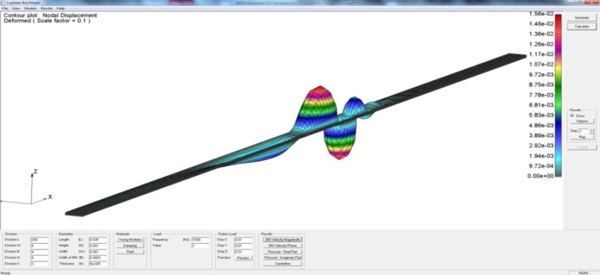
Task 3.1 – BioIRC developed CAD application for generation of the DAT files, which are input files for PAK solver. End user can set geometrical parameters of the cochlea model, set material properties, create mesh, set boundary conditions and loads of the model, either box model or coiled model of the cochlea.
Model can be solved with modal analysis or with time domain analysis.
Obtained results can be seen in CAD application – displacement of the basilar membrane, pressure in the fluid chambers, etc.
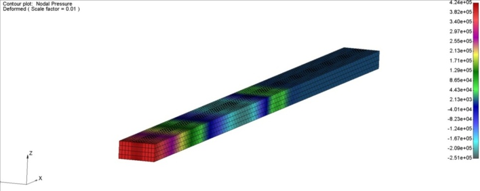
Results for cochlea box model for input frequency of 1 kHz:
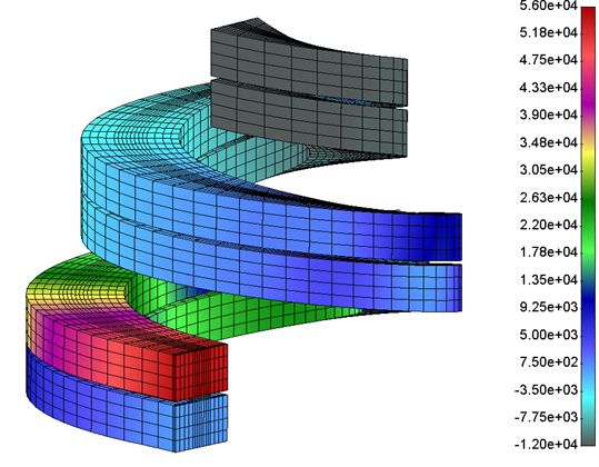
Results for cochlea coiled model for input frequency of 1 kHz:
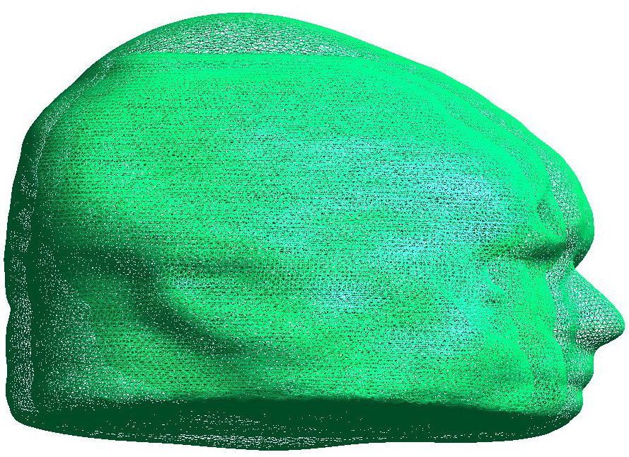
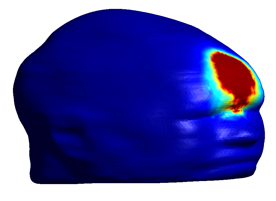
Task 3.3 – BioIRC developed meshing tool for head models, obtained from the CT images. Input files are STL files. Boundary conditions and applied loads are also placed in STL files.
Multiple inputs can be saved in one STL file. Multiple boundary conditions should be saved in one STL file also.
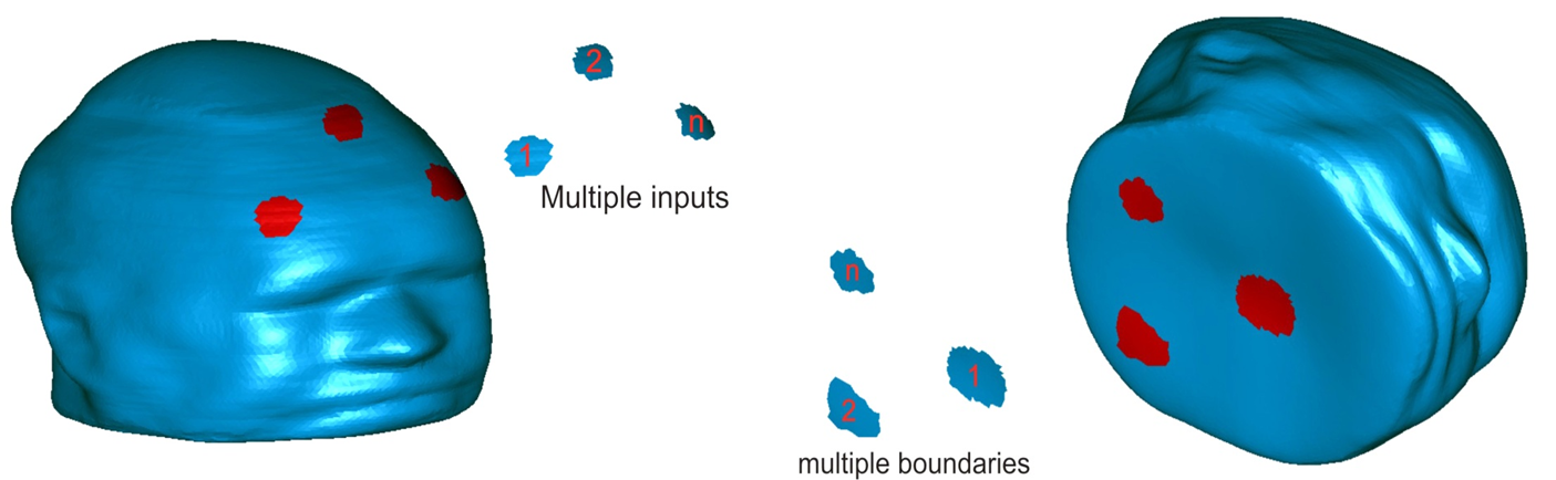
Pre-processing model of the head:
Stress distribution with applied forces in forehead area:
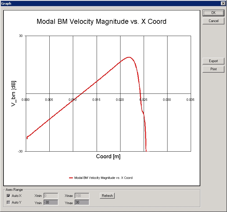
Results - mapping frequency:
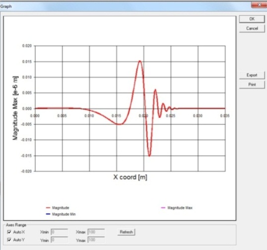
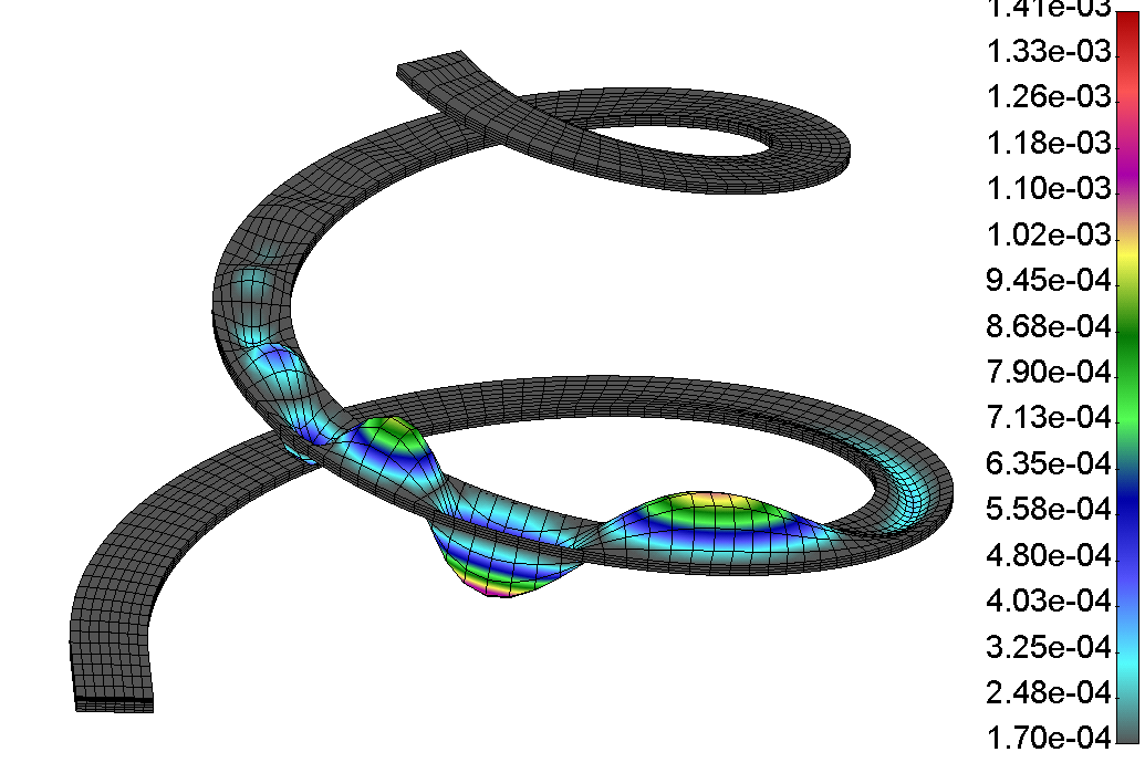
Oscillations of the basilar membrane - spiral model of the cochlea: